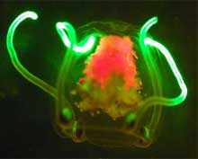
Fig. 1. Small planktonic jellyfish with bright green-fluorescent tentacles. The red fluorescence in the middle of the jellyfish comes from chlorophyll in the ingested algae. Click image for larger view and credits.
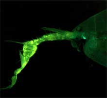
Fig. 2. The front end of the male pontellid copepod possessing enlarged right antenna, the bright fluorescence of which most likely improves the visual mate recognition. Click image for larger view and credits.
Fluorescence: The Secret Color of the Deep
Mikhail V. Matz, PhD
Research Assistant Professor
University of Florida
Fluorescence is a subtle thing. It happens when a fraction of the light illuminating an object is absorbed and then re-emitted as a different color, a two-step process that is necessarily inefficient. In the human visual environment of white daylight, nearly all fluorescent signals get swamped and lost, so special lighting and viewing conditions are required to study them (see more about fluorescence and ways to detect it). Because of this, fluorescence usually does not attract interest of visual ecologists. In the ocean, however, the illumination is predominantly blue (read more about the light in the ocean), so there seems to be a natural opportunity to view fluorescence by unaided eyes: its green, yellow or red color would stand out nicely against the background. We hypothesized that ocean inhabitants would be able to use fluorescence for visual communication, and went out to look for evidence.
It was also a very exciting opportunity to look at the ocean environment with the fluorescence-detecting “alien eye”, to see if we could detect features that thus far went unnoticed. In addition, we are on the lookout for novel fluorescent compounds for biotechnology.
In animals, biologically functional fluorescence is usually bright, concentrated
in certain body parts and shows one or a few well-defined peaks in its
spectrum suggesting involvement of specialized pigments. Thus far, we detected
several examples of this in midwater creatures, which spend all their life
suspended in the “big blue”. In jellyfish and their relatives,
the fluorescence of tentacles and other feeding appendages seems to be
related to prey attraction (Fig. 1, see also the paper about fluorescent lures of a fish-eating jellyfish ![]() ), whereas bright fluorescence in copepods
is clearly for the purpose of visual recognition (Fig. 2). We take these observations
as support for our hypothesis, and are going to continue the search for fluorescence
in midwater animals.
), whereas bright fluorescence in copepods
is clearly for the purpose of visual recognition (Fig. 2). We take these observations
as support for our hypothesis, and are going to continue the search for fluorescence
in midwater animals.
In contrast, fluorescence in the organisms living on the deep sea bottom in the majority of cases seems to be a mere by-product of particular tissue biochemistry and is unlikely to play any particular adaptive role.
Such fluorescence is usually weak, is not localized to particular body
regions and has a wide featureless spectrum. The most common example of
this is green fluorescence of the exoskeleton in crustaceans and other
arthropods such as sea spiders (Fig. 3, see also the ![]() video of the sea spider).
video of the sea spider).
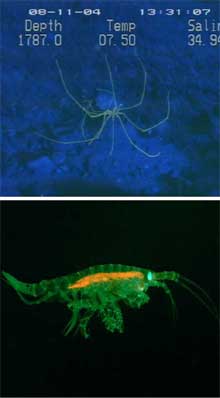
Fig. 3. Fluorescence of a sea spider and a small benthic amphipod. Both the green fluorescence of the whole exoskeleton, as well as weak red of the gut content, are examples of non-functional fluorescence. The cyan fluorescence of the amphipod’s eye is most probably the consequence of visual pigment presence and is also unlikely to have any specific biological function. Click image for larger view and image credit.
The reason for such a trend may be that the vision capabilities of bottom-dwellers
are on average much less developed that in midwater organisms, so there
is simply nobody to see the super-weak fluorescent signals that might be
spawned by the tiny amounts of remaining downwelling light and bioluminescence
of the local fauna (read more about bioluminescence in the deep sea ![]() ). The situation may be quite different in shallow water
where plenty of light is available (see the report of fluorescent signaling in mantis shrimp
). The situation may be quite different in shallow water
where plenty of light is available (see the report of fluorescent signaling in mantis shrimp ![]() ). In addition, last year we found two remarkable exceptions from the rule – tube
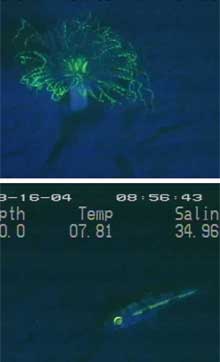
anemone and a fish, both deep bottom-dwelling animals that clearly possess fluorescence “on purpose” (Fig. 4, see also the
). In addition, last year we found two remarkable exceptions from the rule – tube
anemone and a fish, both deep bottom-dwelling animals that clearly possess fluorescence “on purpose” (Fig. 4, see also the ![]() video of the fish).
The function of fluorescence in these creatures is a total mystery thus
far, which we plan to address in the future studies.
video of the fish).
The function of fluorescence in these creatures is a total mystery thus
far, which we plan to address in the future studies.
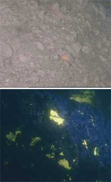
Fig. 5. Appearance of methane hydrates on the sea floor in the white light (top) and through fluorescent optics (bottom). Click image for larger view and credits.
Finally, just looking around with our “alien eye” resulted
in one very exciting observation last year: methane hydrates (“methane ice ![]()
![]() ”), which, at least at the site where we observed them, turned out to be extremely
brightly fluorescent, although they were quite inconspicuous under normal lighting
conditions (Fig. 5). Methane hydrates, in addition to being a potential energy
source, were suggested to have played a critical role in climate change
”), which, at least at the site where we observed them, turned out to be extremely
brightly fluorescent, although they were quite inconspicuous under normal lighting
conditions (Fig. 5). Methane hydrates, in addition to being a potential energy
source, were suggested to have played a critical role in climate change ![]() .
Our observation may lead to a new approach for mapping them on the seafloor;
however, more observations are required to develop a proper technology.
.
Our observation may lead to a new approach for mapping them on the seafloor;
however, more observations are required to develop a proper technology.


