Measuring Vision in Crustaceans
Tammy FrankAssociate Scientist
Harbor Branch Oceanographic Institution
How do you study the visual system of a crustacean – in this case, a shrimp or a crab?
Scientists will first remove the specimen's eyes, and then prepare them under dim red light for structural studies. Using both transmission and scanning electron microscopy, the studies will indicate whether any unusual optical adaptations significantly increase the animal's sensitivity to light. Past research indicates that the eyes of deep-sea organisms have wide variety of unusual adaptations: biological light guides, scanning binoculars, huge parabolic mirrors, and vast arrays of reflecting mirrors behind the retina.
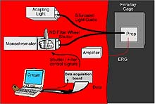
We will also complete physiological studies to determine what colors the samples see, and how sensitive their eyes are compared to shallow-water species. Since a shrimp or crab cannot communicate, information is gathered directly from its eye. In order to do this, we assemble a complex equipment system at sea (Figure 1), and secure the system for heavy seas.

Figure 1: A diagram of the equipment needed to measure the response of an eye to a flash of light. Click image for larger view.
Equipment for Measuring Vision
Measuring vision requires many pieces of equipment. The shrimp or crab is placed in a holder that allows its pleopods (legs, in the shrimp) or gill bailers (in the crab) to move and generate respiratory currents across its gills. The specimen is kept in a light-tight room, in a bath of chilled water, with an electrode on one eye. When the eye sees a flash of light, it converts the light energy into electrical energy, which is picked up by the electrode on the eye. This electrical signal is amplified and stored as a digitized signal on the computer.
The monochromator, a device that splits white light into its component colors, allows scientists to select the color of light to be flashed on the sample. A neutral density filter wheel, located on the front of the monochromator, allows the scientist to control the intensity of the light. The Faraday cage blocks out electrical noise that the electrode could pick up. A light guide is used to transmit the light from the monochromator (and not the room) to the eye. One end of the light guide is sealed to the monochromator. The other end goes through a little hole in the Faraday cage and transmits the light directly to the eye. Based on the size of the response that the eye gives to a flash of light, the scientist can tell what colors the it sees the best. For example, if the eye is most sensitive to blue light, the measured electrical response to blue light will be much bigger than the response to red light of the same intensity

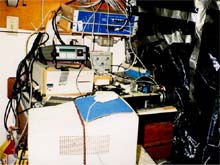
Figure 2: This is what the lab really looks like at sea. On a ship, you have to fit into whatever space is available, building light-tight rooms out of black plastic and duct tape. Click image for larger view.
Continuous Motion?
Another set of experiments will indicate the critical flicker fusion frequency (CFF) in the crustaceans. This is the frequency at which a flickering light source is considered continuous.
A good way to understand CFF is to think about movies and how they work. Modern movies, for example, are actually individual snapshots presented so rapidly that you can’t see the flicker. Early movies, on the other hand, showed 16 frames per second, which is considerably slower than the human flicker fusion frequency, so people are able to detect the flicker between frames.
That's why the motion of people walking in these early movies looks halting and spastic. Today, the rate is 24 frames per second, but each frame is flashed 3 times, increasing the flicker rate to 72 flashes per second -- above our CFF -- so the motion of people walking looks fluid and continuous. A crustacean isn’t interested in watching movies, but it does need to be able to track its prey. The CFF is an indication of how well the crustacean is able to do that.


