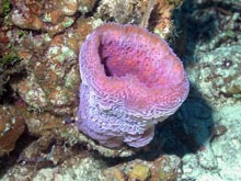
Some sponges, like this vase sponge (Callyspongia plicifera), do not host a microbial community that is substantially different from that found in the surrounding water column. Other sponges contain microorganisms that are not commonly found in the water, and they are considered bacteriosponges. Click image for larger view and image credit.
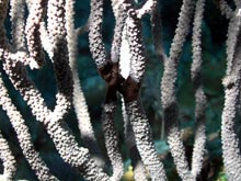
In general, most microorganisms are too small to be seen with the naked eye. However, these tufts of filamentous cyanobacteria form large accumulations of individual microscopic cells that are visible. Cyanobacteria often form large aggregations that produce toxins and are detrimental to marine organisms and humans. Collectively, we call these aggregations "harmful algal blooms." Click image for larger view and image credit.
The Ocean’s Invisible Majority
May 25, 2007
Julie Olson
University of Alabama
![]() The many steps required to process microbial samples.
(Quicktime, 7.1 Mb.)
The many steps required to process microbial samples.
(Quicktime, 7.1 Mb.)
Although not seen by the naked eye, a milliliter (1/1000 of a liter) of seawater contains thousands to millions of bacterial cells. Marine surfaces, ranging from sand particles to corals, also contain large numbers of microorganisms. These tiny animals play many roles in the marine environment, including serving as food for filter-feeding organisms, cycling nutrients, and causing diseases. As with humans, marine organisms are often colonized by microbes, usually with no apparent negative impacts and quite likely with benefits (e.g., protective normal flora preventing pathogens from attaching, nutrient cycling). In some cases, the normal balance is disrupted and pathogenic microorganisms become established, resulting in disease. The recent increase in the reported decline of coral reefs has been largely attributed to coral diseases, although relatively few pathogens have been identified.
Many sponges are considered to be bacteriosponges as they contain large numbers of bacteria inside the sponge tissues. Up to 40% of the "sponge" biomass can actually consist of microorganisms. These sponges do not appear to be harmed by these bacterial associations, and a number of potential reasons for their existence have been proposed, including nutrient acquisition and movement inside the organism, stabilization of the sponge skeleton, and provision of chemical defenses. Some of these sponges contain compounds, whether of sponge or microbial origin, which appear to deter predation and also may be useful as human pharmaceutical products.
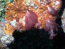
Shallow-water corals are also associated with a diverse microflora, including photosynthetic dinoflagellates called "zooxanthellae," which are responsible for carrying out photosynthesis within coral tissues. These small organisms provide a source of carbon for their host coral and give the coral its distinctive coloration. In deeper waters where no light penetrates, corals have been found that do not have zooxanthellae and instead rely on particles (and bacteria) within the water for their carbon requirements. The surface of coral tissue often has a thick slime layer, which is colonized by a large diversity of microorganisms. As the mucus sloughs off of the host coral, the microorganisms embedded in this matrix are released into the water column.
Thus, the marine environment supports a very large and diverse community of microorganisms. Relatively little is known about the functions of marine microbes and their interactions, both beneficial and harmful, with other organisms. Information about microbial community structure across a depth gradient, such as we will be working with on our wall site, will be extremely useful for determining the potential roles of associated microorganisms.

Cyanobacteria are increasingly common on Caribbean coral reefs and are frequently found overgrowing benthic (bottom-dwelling) organisms, such as this soft coral (Eunicea sp.). Although some cyanobacteria form symbiotic relationships with a host organism, others are detrimental and can cause tissue necrosis and mortality. Click image for larger view and image credit.

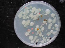
Each spot on this agar plate is a bacterial colony recovered from a marine sponge sample. By the time a colony is visible to the human eye, it consists of at least one million cells. Less than 1% of bacteria can be grown under laboratory conditions, suggesting that there is a large reservoir of unknown microbial diversity. As a result, microbiologists use a combination of cultivation and molecular genetic characterizations to examine microbial communities. Click image for larger view and image credit.


