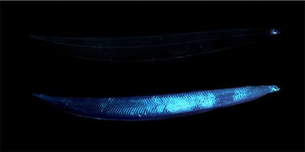
Two images of the same leptocephalus eel larva. The top image is viewed under unpolarized, transmitted light. The bottom image is viewed under polarized, transmitted light by a camera with a polarizing filter. The increased visibility of the bottom image is due to the presence of birefringent muscle and connective tissue fibers. Photo composite: S Johnsen using images from E. Widder.
Related Links


